
Friday, June 24, 2016
Wednesday, June 22, 2016

Sunday, June 19, 2016


Saturday, June 18, 2016
Otoscopy
The procedure for otoscopy is as :
Introduce yourself.
Tell that the chart has been reviewed and examination done, and i further need to examine ears for making a final diagnosis.
Explain the procedure steps to Child and his parents.
Tell them and show the device that it just emits a light and with light the ear will be seen . it causes no pain . Throw light on the hand of the child and talk with him to build reputation.
For a very small child the position is the same as holding in LAP for examining the throat. for elder child ask mother to stand beside and and have her hand on his head so as he doesnt move it.
Then inspect the Pinna , Behind the ear and the mastoid process .
then with Right sized speculum on the auriscope , hold the scope like a PEN , examine the external auditory canal for WAX , REDNESS SWELLING DISCHARGE, then the pearly white eardrum, the cone of light that forms as light is thrown over it and the fluid level seen through it if any, or the perforation. With a bladder attached to the scope check movements of the eardrum. Withdraw the scope, examine the other ear .
Tell findings to the parents ,
Greet and good bye.
Write findings in the chart.
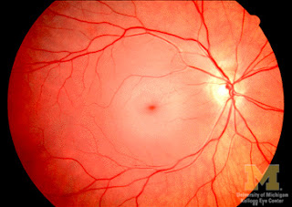
Fundoscopy
Technique of Fundoscopy.
First I shall introduce myself to the patient/child and his parents.
Then tell them that i have reviewed the Chart and done physical examination and i further need to examine the eyes. Tell them that its a simple device that illuminates the inside of the eye and no pain or harm will be done to the child. Make reputation with the child and show him the light fall on his hand .
Then Instill Tropicamide eye drops for Dilatation of pupil and Atropine eye drops for Cyclopegic effect.
Sit at level with the child at 1 arm length.
Right eye of examiner for right eye of patient.
Left eye of examiner for left eye of patient.
Ask the patient to see distant object and hold fundoscope about 1 arm length and move closer till you see the RED REFLEX. RED reflex denotes that the media of the eye from The cornea to Lens to Vitreous humor is clear . Keeping Red Reflex in focus move closer and closer to the eye till blood vessels are visible , now as one vessel is traced move Nasally where all vessels are converging , it is the Optic Disc. It is Pale to pink in colour with a greyish round margin. papilledema, the vessels , their tortuosity if any , hemorrhages on the retina, chery red spot are observed. Papilledema has 5 grades,
grade 1 half of the disc is edematous
grade two circumferential edema of disc
grade 3 disc edema + vessels as they move away from the disc become tortuous and appear 'broken'
grade 4 vessels appear tortuous and broken just as they emerge from the disc
grade 5 no clear demarcation of the disc.
Fundoscopy may also help identify if the eye is Emmetropic, myopic, or hypermetropic.
Examine the other eye.
Write notes of the findings in the chart.
Inform parents about the findings and further steps to be taken accordingly.
Ask patient to not to drive till the effect of medicine goes away, and ask attendants to help patient walk about as he cannot see clearly.
Greet and Good bye.
 CHERY RED SPOT
A cherry-red spot is a finding in the macula of the eye in a variety of lipid storage disorders and in central retinal artery occlusion. It describes the appearance of a small circular choroid shape as seen through the fovea centralis. Its appearance is due to a relative transparency of the macula; storage disorders cause the accumulation of storage material within the cell layers of the retina, however, the macula, which is relatively devoid of cellular layers, does not build up this material, and thus allows the eye to see through the macula to the red choroid below.
Chery red spot may be seen in TAY SACHS disease, hurlers disease, Nieman picks diseases , CO poisoning , Quinine toxicity, metachromatic leukodystrophy.
First I shall introduce myself to the patient/child and his parents.
Then tell them that i have reviewed the Chart and done physical examination and i further need to examine the eyes. Tell them that its a simple device that illuminates the inside of the eye and no pain or harm will be done to the child. Make reputation with the child and show him the light fall on his hand .
Then Instill Tropicamide eye drops for Dilatation of pupil and Atropine eye drops for Cyclopegic effect.
Sit at level with the child at 1 arm length.
Right eye of examiner for right eye of patient.
Left eye of examiner for left eye of patient.
Ask the patient to see distant object and hold fundoscope about 1 arm length and move closer till you see the RED REFLEX. RED reflex denotes that the media of the eye from The cornea to Lens to Vitreous humor is clear . Keeping Red Reflex in focus move closer and closer to the eye till blood vessels are visible , now as one vessel is traced move Nasally where all vessels are converging , it is the Optic Disc. It is Pale to pink in colour with a greyish round margin. papilledema, the vessels , their tortuosity if any , hemorrhages on the retina, chery red spot are observed. Papilledema has 5 grades,
grade 1 half of the disc is edematous
grade two circumferential edema of disc
grade 3 disc edema + vessels as they move away from the disc become tortuous and appear 'broken'
grade 4 vessels appear tortuous and broken just as they emerge from the disc
grade 5 no clear demarcation of the disc.
Fundoscopy may also help identify if the eye is Emmetropic, myopic, or hypermetropic.
Examine the other eye.
Write notes of the findings in the chart.
Inform parents about the findings and further steps to be taken accordingly.
Ask patient to not to drive till the effect of medicine goes away, and ask attendants to help patient walk about as he cannot see clearly.
Greet and Good bye.

A cherry-red spot is a finding in the macula of the eye in a variety of lipid storage disorders and in central retinal artery occlusion. It describes the appearance of a small circular choroid shape as seen through the fovea centralis. Its appearance is due to a relative transparency of the macula; storage disorders cause the accumulation of storage material within the cell layers of the retina, however, the macula, which is relatively devoid of cellular layers, does not build up this material, and thus allows the eye to see through the macula to the red choroid below.
Chery red spot may be seen in TAY SACHS disease, hurlers disease, Nieman picks diseases , CO poisoning , Quinine toxicity, metachromatic leukodystrophy.


Thursday, June 16, 2016


Wednesday, June 15, 2016


Monday, June 13, 2016


Sunday, June 12, 2016

Subscribe to:
Posts (Atom)
-
 tags: histology slides histology diagrams kemu human histology (c) www.Tsnaps.tk
 -
 histological diagram of transverse section of trachea trachea histology slide Also visit : http://histology-slides-database.blogspot.c...
 -
 lymph node histology slide Click to enlarge the image tags : Histology Of lymph node Histology Slide Of lymph node Histological Slide o...