
Monday, April 30, 2018

Chest examination (and relevant) steps / respiratory system examination
These are steps for CHEST and relevant examination.
If asked to examine respiratory system , then it begins from examination of nose , ears , and oral cavity .................
If asked to examine respiratory system , then it begins from examination of nose , ears , and oral cavity .................

Sunday, April 29, 2018
DSD
Denys Drash
Phenotypic female or male
46xy
Female internal organs with streak ovaries(as MIF is missing)(WT1 suppressor defect)
Plus syndrome findings wilms,renal failure,ambiguous genitals.
Abdominal mass is a rare presenting symptom, they re actually treated as atypical nephrotics ;)
Because genitalia may be virilized to be male looking with labial fusions. If male type true genitals,then cryptorchidism.,but still female internal organs present(obviously has MIF was not there so internal organs will form).
WAGR
phenotypic male but with cryptorchdism and hypospadias.
46 XY
Wilms,aniridia,GU abnor,retard mental
Defect in chromosome 11.
If the patient has all features of..... DenysDrash,but wilms tunor is not there(oops), then look for developing Gonadoblastoma in streak gonads,its Frasier syndrome.
Now theres a female,
Who has failed to achieve menarche,
But there is thelarche,
Genitals female type,
Ultrasound reveals that there are no internal female organs, and along tract,instead, testes are seen.
Means MIF worked, so no internal organs, testosterons didnot work thats why female external organs. Thats androgen insensitivity syndrome.
Her karyoype will be 46xy.,and normal testosterone...that converts to estradiol,and so thelarche is there.
Now, after this patient,
Another female comes :)
She has failed to achieve menarche, but his patient, also doesnt have thelarche,
The genitals are true female type,
And ultrasound reveals, internal female organs n gonads....oppps! Wait n see???
No.
Get Karyotype.
.
46xy
.
Get testosterone.
.
Low.
.
.thats defective SRY gene,
46,xy but no SRY, means No MIF, means female genitals will form,
No SRY means no testosterone,
Which means no male ductal system or testes and so female genitals.
Its Swyer syndrome.
Saturday, April 28, 2018

Thursday, April 26, 2018
Self
*Imam Ibn al-Qayyim* (rahimahullah):
A friend will not (literally) share your struggles, and a loved one cannot physically take away your pain, and a close one will not stay up the night on your behalf. So look after yourself, protect yourself, nurture yourself and don't give life's events more than what they are really worth.
Know for certain that when you break no one will heal you except you, and when you are defeated no one will give you victory except your determination. Your ability to stand up again and carry on is your responsibility.
Do not look for your self worth in the eyes of people; look for your worth from within your conscious. If your conscious is at peace then you will ascend high and if you truly know yourself then what is said about you won't harm you .
Do not carry the worries of this life because this is for Allah. And do not carry the worries of sustenance because it is from Allah. And do not carry the anxiety for the future because it is in the Hands of Allah.
Carry one thing: How to Please Allah. Because if you please Him, He pleases you, fulfils you and enriches you like.
Do not weep from a life that made your heart weep. Just say, "Oh Allah compensate me with good in this life and the HEREAFTER."
Sadness departs with a Sajdah. Happiness comes with a sincere Du'a. Allah does not forget the good you do. Nor does he forget the good you did to others and the pain you relieved them from. Nor will He forget the eye which was about to cry but you made it laugh.
Live your life with this principle:
*Be good even if you don't receive good, not because of others' sake but because Allah loves those who do good*
Wednesday, April 25, 2018
Fundus and Retinal Photography using cell phone camera
The first experience of using a direct fundoscope is tedious but if successful, it's rewarding.Because apart from the importance of clinical signs in retina/fundus, we find that retina is an elegant tissue. Behind the lens there is big world of vessels, and a world of clinical signs. No doubt eye is a window to brain, but to see through this window, we need a good practice with the use of direct opthalmoscope, and patience if our patient is non-cooperative(specially kids). Indirect opthalmoscopy may be easier. However, the instrument for (bedside) fundoscopy is quite complex in its optics, and needs good expertise to use it rapidly to see retina.
 During my house job rotation in East Medical Ward, Mayo Hospital,Lhe, my seniors introduced me to this instrument, direct opthalmoscope. The model they carried back then was the same one as in photograph above.
With simple instructions given by them, after an effort of 2 minutes , i was able to see just a glimpse of vessels in retina. It was all very beautiful. But i could not locate fundus or track vessels.
Very few doctors carried the instrument with them as it was ëxpensive. And so, everyone relied on eye department for trivial retinal findings.
The physics that is involved in opthalmoscope is a really simple one. I illustrate it here as :

The rays of light from bulb(a) get collimated(parallel) by lens(b) , mirror(c) deflects them 90 degrees towards patient's eye which then go through patient's pupil>lens>strike retina > return from retina>lens>pupil and come back just near to mirror of ophtalmoscope, and physician sees those light rays through aperture(g).
From this physics, i ascertained the issue, an issue that"direct opthalmoscopy is hard to learn ,and not everyone can do it in the first go".
I guessed that the distance "i" is making the job tedious.
The second issue , "expensive" instrument, was because of the optics used in the instrument.
To eliminate all optics, i came up with an idea, to use Surface Mount Device type Light Emitting Diode as a light source, on a piece of card or preferably Printed Circuit Board, and make an aperture in it so that the distance "i"is considerably reduced.
I also found out that , if SMD LED is placed close to cell phone camera , very close , then cellphone camera works like a fundoscope. If you understand photography, then you know that FLASH is placed a bit far from Camera so that RED EYE REFLEX is minimized. I brought LED closer to camera, to make RED REFLEX more pronounced , and once very close to eye, to see through eye.
Follow below.


The above two diagrams illustrate well the idea. Distance "i"is considerably reduced.
The initial version made according to the plan looked very much like this :


This device was named as OptiCard, it had an aperture in its upper piece for direct fundoscopy (without cell phone camera), and the device could be attached to cellphone camera by folding the upper piece of card, as below :

The image of retina visualized using cellphone :

By reducing the distance "i", everyone can do fundoscopy in the first go.
More research data is needed however, to prove my point.
(I have shared the link to my research paper below).
So the instrument as per end of year 2017 , looked very much similar to the one in above photos.
It had two pieces.
In 2018,
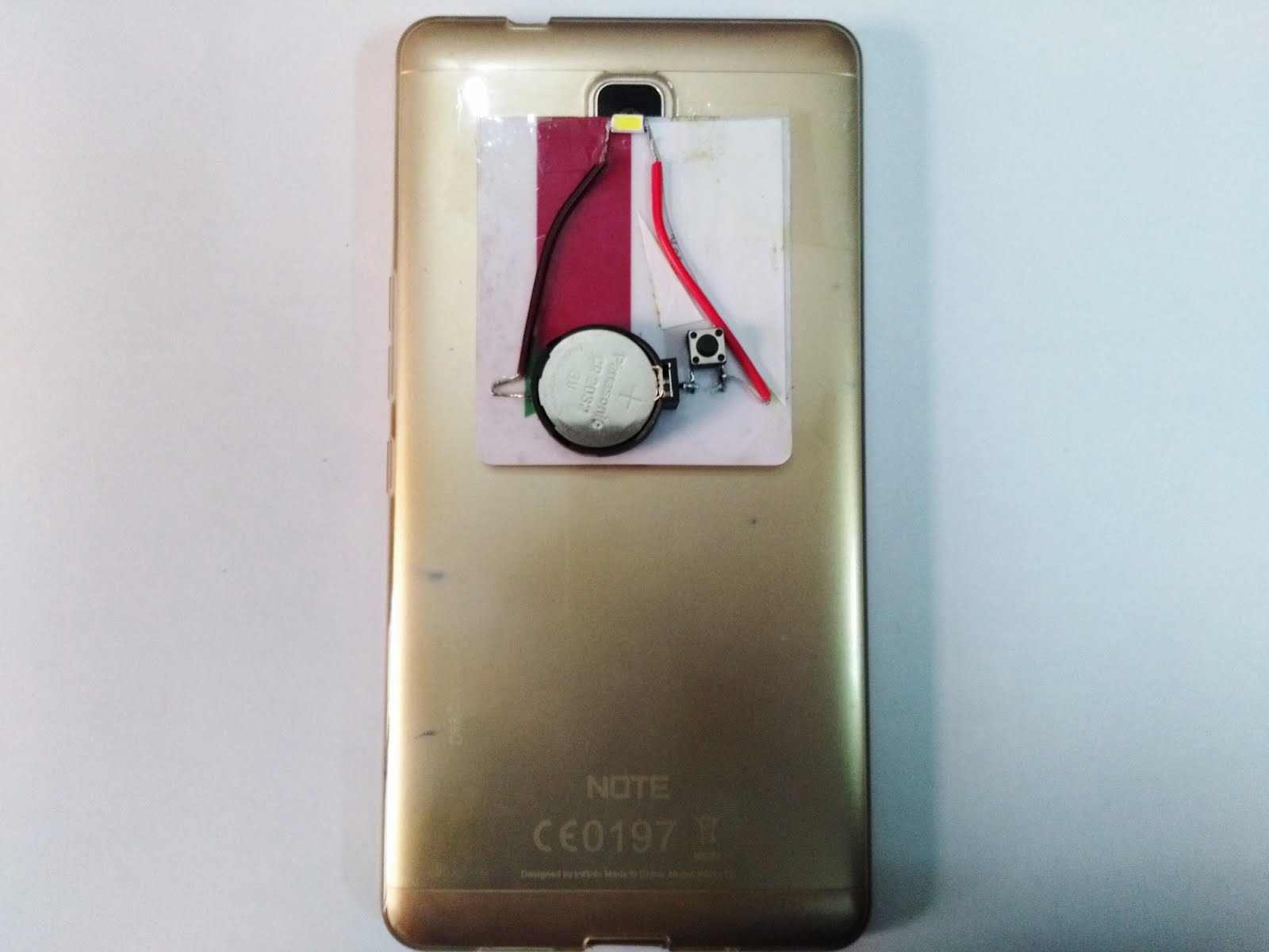
I came up with another idea, to make aperture in the card below LED.
I used printed circuit board, and SMD switch and the final device looks like :
 Front
Front
 Back
Back
 Attached to cellphone Camera

The video demonstration of the gadget is as :
With the intent to create a less expensive device I found an easier way too , to visualize retina, a very simple device, getting rid of complex(& expensive optics).
Dr M. Tauseef Omer
FCPS Resident
Dept of Paediatric Medicine & Neonatology
MAYO Hospital,King Edward Medical University,
Lahore, Pakistan.
contact@tlabs.pk
tauseef_ravian153@hotmail.com
www.tlabs.pk
 During my house job rotation in East Medical Ward, Mayo Hospital,Lhe, my seniors introduced me to this instrument, direct opthalmoscope. The model they carried back then was the same one as in photograph above.
With simple instructions given by them, after an effort of 2 minutes , i was able to see just a glimpse of vessels in retina. It was all very beautiful. But i could not locate fundus or track vessels.
Very few doctors carried the instrument with them as it was ëxpensive. And so, everyone relied on eye department for trivial retinal findings.
The physics that is involved in opthalmoscope is a really simple one. I illustrate it here as :

The rays of light from bulb(a) get collimated(parallel) by lens(b) , mirror(c) deflects them 90 degrees towards patient's eye which then go through patient's pupil>lens>strike retina > return from retina>lens>pupil and come back just near to mirror of ophtalmoscope, and physician sees those light rays through aperture(g).
From this physics, i ascertained the issue, an issue that"direct opthalmoscopy is hard to learn ,and not everyone can do it in the first go".
I guessed that the distance "i" is making the job tedious.
The second issue , "expensive" instrument, was because of the optics used in the instrument.
To eliminate all optics, i came up with an idea, to use Surface Mount Device type Light Emitting Diode as a light source, on a piece of card or preferably Printed Circuit Board, and make an aperture in it so that the distance "i"is considerably reduced.
I also found out that , if SMD LED is placed close to cell phone camera , very close , then cellphone camera works like a fundoscope. If you understand photography, then you know that FLASH is placed a bit far from Camera so that RED EYE REFLEX is minimized. I brought LED closer to camera, to make RED REFLEX more pronounced , and once very close to eye, to see through eye.
Follow below.


The above two diagrams illustrate well the idea. Distance "i"is considerably reduced.
The initial version made according to the plan looked very much like this :


This device was named as OptiCard, it had an aperture in its upper piece for direct fundoscopy (without cell phone camera), and the device could be attached to cellphone camera by folding the upper piece of card, as below :
The image of retina visualized using cellphone :

By reducing the distance "i", everyone can do fundoscopy in the first go.
More research data is needed however, to prove my point.
(I have shared the link to my research paper below).
So the instrument as per end of year 2017 , looked very much similar to the one in above photos.
It had two pieces.
In 2018,
I came up with another idea, to make aperture in the card below LED.
I used printed circuit board, and SMD switch and the final device looks like :
 Front
Front Back
Back Attached to cellphone Camera
The video demonstration of the gadget is as :
The research paper published in Journal of College of Physicians and Surgeons Pakistan (JCPSP) is here:
The project is still ongoing, and I have multiple updates in my mind.
I have started collecting a digital record of retinal findings of patients , the "Photo FollowUp".With the intent to create a less expensive device I found an easier way too , to visualize retina, a very simple device, getting rid of complex(& expensive optics).
Dr M. Tauseef Omer
FCPS Resident
Dept of Paediatric Medicine & Neonatology
MAYO Hospital,King Edward Medical University,
Lahore, Pakistan.
contact@tlabs.pk
tauseef_ravian153@hotmail.com
www.tlabs.pk
Friday, April 20, 2018
Sturge weber syndrome
Sturge Weber syndrome also known as sturge Weber Dimitri syndrome or encephalo Trigeminal angiomatosis it has port wine stain which is a facial hemangioma classical e in the distribution of the first division of fifth cranial nerve that is Trigeminal nerve the patient has focal seizures on the contralateral size and hemangiomatous changes of the meninges on the ipsilateral side as port wine stain and so calcifications develop on the ipsilateral side which are seen on the CT scan and gyriform form calcifications sturge Weber syndrome or encephalo Trigeminal angiomatosis is a congenital disease it has no genetic basis its fits are very difficult to control there may be associated contralateral hemiparesis and intellectual disability
Subscribe to:
Posts (Atom)
-
 tags: histology slides histology diagrams kemu human histology (c) www.Tsnaps.tk
 -
 histological diagram of transverse section of trachea trachea histology slide Also visit : http://histology-slides-database.blogspot.c...
 -
 lymph node histology slide Click to enlarge the image tags : Histology Of lymph node Histology Slide Of lymph node Histological Slide o...