
Monday, April 30, 2018
Chest examination (and relevant) steps / respiratory system examination
These are steps for CHEST and relevant examination.
If asked to examine respiratory system , then it begins from examination of nose , ears , and oral cavity .................
If asked to examine respiratory system , then it begins from examination of nose , ears , and oral cavity .................

Sunday, April 29, 2018
DSD
Denys Drash
Phenotypic female or male
46xy
Female internal organs with streak ovaries(as MIF is missing)(WT1 suppressor defect)
Plus syndrome findings wilms,renal failure,ambiguous genitals.
Abdominal mass is a rare presenting symptom, they re actually treated as atypical nephrotics ;)
Because genitalia may be virilized to be male looking with labial fusions. If male type true genitals,then cryptorchidism.,but still female internal organs present(obviously has MIF was not there so internal organs will form).
WAGR
phenotypic male but with cryptorchdism and hypospadias.
46 XY
Wilms,aniridia,GU abnor,retard mental
Defect in chromosome 11.
If the patient has all features of..... DenysDrash,but wilms tunor is not there(oops), then look for developing Gonadoblastoma in streak gonads,its Frasier syndrome.
Now theres a female,
Who has failed to achieve menarche,
But there is thelarche,
Genitals female type,
Ultrasound reveals that there are no internal female organs, and along tract,instead, testes are seen.
Means MIF worked, so no internal organs, testosterons didnot work thats why female external organs. Thats androgen insensitivity syndrome.
Her karyoype will be 46xy.,and normal testosterone...that converts to estradiol,and so thelarche is there.
Now, after this patient,
Another female comes :)
She has failed to achieve menarche, but his patient, also doesnt have thelarche,
The genitals are true female type,
And ultrasound reveals, internal female organs n gonads....oppps! Wait n see???
No.
Get Karyotype.
.
46xy
.
Get testosterone.
.
Low.
.
.thats defective SRY gene,
46,xy but no SRY, means No MIF, means female genitals will form,
No SRY means no testosterone,
Which means no male ductal system or testes and so female genitals.
Its Swyer syndrome.
Saturday, April 28, 2018

Thursday, April 26, 2018
Self
*Imam Ibn al-Qayyim* (rahimahullah):
A friend will not (literally) share your struggles, and a loved one cannot physically take away your pain, and a close one will not stay up the night on your behalf. So look after yourself, protect yourself, nurture yourself and don't give life's events more than what they are really worth.
Know for certain that when you break no one will heal you except you, and when you are defeated no one will give you victory except your determination. Your ability to stand up again and carry on is your responsibility.
Do not look for your self worth in the eyes of people; look for your worth from within your conscious. If your conscious is at peace then you will ascend high and if you truly know yourself then what is said about you won't harm you .
Do not carry the worries of this life because this is for Allah. And do not carry the worries of sustenance because it is from Allah. And do not carry the anxiety for the future because it is in the Hands of Allah.
Carry one thing: How to Please Allah. Because if you please Him, He pleases you, fulfils you and enriches you like.
Do not weep from a life that made your heart weep. Just say, "Oh Allah compensate me with good in this life and the HEREAFTER."
Sadness departs with a Sajdah. Happiness comes with a sincere Du'a. Allah does not forget the good you do. Nor does he forget the good you did to others and the pain you relieved them from. Nor will He forget the eye which was about to cry but you made it laugh.
Live your life with this principle:
*Be good even if you don't receive good, not because of others' sake but because Allah loves those who do good*
Wednesday, April 25, 2018
Fundus and Retinal Photography using cell phone camera
The first experience of using a direct fundoscope is tedious but if successful, it's rewarding.Because apart from the importance of clinical signs in retina/fundus, we find that retina is an elegant tissue. Behind the lens there is big world of vessels, and a world of clinical signs. No doubt eye is a window to brain, but to see through this window, we need a good practice with the use of direct opthalmoscope, and patience if our patient is non-cooperative(specially kids). Indirect opthalmoscopy may be easier. However, the instrument for (bedside) fundoscopy is quite complex in its optics, and needs good expertise to use it rapidly to see retina.
 During my house job rotation in East Medical Ward, Mayo Hospital,Lhe, my seniors introduced me to this instrument, direct opthalmoscope. The model they carried back then was the same one as in photograph above.
With simple instructions given by them, after an effort of 2 minutes , i was able to see just a glimpse of vessels in retina. It was all very beautiful. But i could not locate fundus or track vessels.
Very few doctors carried the instrument with them as it was ëxpensive. And so, everyone relied on eye department for trivial retinal findings.
The physics that is involved in opthalmoscope is a really simple one. I illustrate it here as :

The rays of light from bulb(a) get collimated(parallel) by lens(b) , mirror(c) deflects them 90 degrees towards patient's eye which then go through patient's pupil>lens>strike retina > return from retina>lens>pupil and come back just near to mirror of ophtalmoscope, and physician sees those light rays through aperture(g).
From this physics, i ascertained the issue, an issue that"direct opthalmoscopy is hard to learn ,and not everyone can do it in the first go".
I guessed that the distance "i" is making the job tedious.
The second issue , "expensive" instrument, was because of the optics used in the instrument.
To eliminate all optics, i came up with an idea, to use Surface Mount Device type Light Emitting Diode as a light source, on a piece of card or preferably Printed Circuit Board, and make an aperture in it so that the distance "i"is considerably reduced.
I also found out that , if SMD LED is placed close to cell phone camera , very close , then cellphone camera works like a fundoscope. If you understand photography, then you know that FLASH is placed a bit far from Camera so that RED EYE REFLEX is minimized. I brought LED closer to camera, to make RED REFLEX more pronounced , and once very close to eye, to see through eye.
Follow below.


The above two diagrams illustrate well the idea. Distance "i"is considerably reduced.
The initial version made according to the plan looked very much like this :


This device was named as OptiCard, it had an aperture in its upper piece for direct fundoscopy (without cell phone camera), and the device could be attached to cellphone camera by folding the upper piece of card, as below :

The image of retina visualized using cellphone :

By reducing the distance "i", everyone can do fundoscopy in the first go.
More research data is needed however, to prove my point.
(I have shared the link to my research paper below).
So the instrument as per end of year 2017 , looked very much similar to the one in above photos.
It had two pieces.
In 2018,
I came up with another idea, to make aperture in the card below LED.
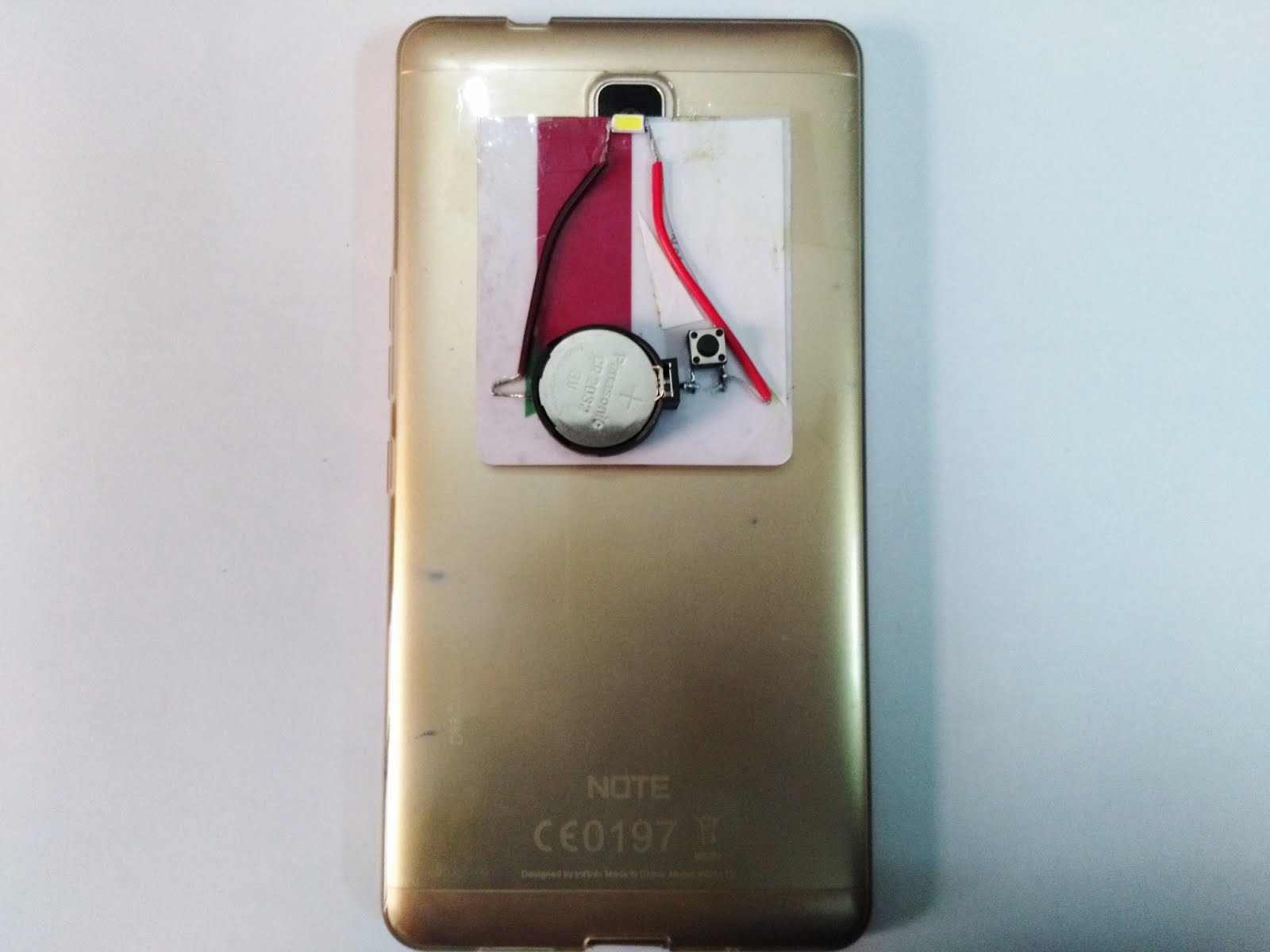
I used printed circuit board, and SMD switch and the final device looks like :
 Front
Front
 Back
Back
 Attached to cellphone Camera

The video demonstration of the gadget is as :
With the intent to create a less expensive device I found an easier way too , to visualize retina, a very simple device, getting rid of complex(& expensive optics).
Dr M. Tauseef Omer
FCPS Resident
Dept of Paediatric Medicine & Neonatology
MAYO Hospital,King Edward Medical University,
Lahore, Pakistan.
contact@tlabs.pk
tauseef_ravian153@hotmail.com
www.tlabs.pk
 During my house job rotation in East Medical Ward, Mayo Hospital,Lhe, my seniors introduced me to this instrument, direct opthalmoscope. The model they carried back then was the same one as in photograph above.
With simple instructions given by them, after an effort of 2 minutes , i was able to see just a glimpse of vessels in retina. It was all very beautiful. But i could not locate fundus or track vessels.
Very few doctors carried the instrument with them as it was ëxpensive. And so, everyone relied on eye department for trivial retinal findings.
The physics that is involved in opthalmoscope is a really simple one. I illustrate it here as :

The rays of light from bulb(a) get collimated(parallel) by lens(b) , mirror(c) deflects them 90 degrees towards patient's eye which then go through patient's pupil>lens>strike retina > return from retina>lens>pupil and come back just near to mirror of ophtalmoscope, and physician sees those light rays through aperture(g).
From this physics, i ascertained the issue, an issue that"direct opthalmoscopy is hard to learn ,and not everyone can do it in the first go".
I guessed that the distance "i" is making the job tedious.
The second issue , "expensive" instrument, was because of the optics used in the instrument.
To eliminate all optics, i came up with an idea, to use Surface Mount Device type Light Emitting Diode as a light source, on a piece of card or preferably Printed Circuit Board, and make an aperture in it so that the distance "i"is considerably reduced.
I also found out that , if SMD LED is placed close to cell phone camera , very close , then cellphone camera works like a fundoscope. If you understand photography, then you know that FLASH is placed a bit far from Camera so that RED EYE REFLEX is minimized. I brought LED closer to camera, to make RED REFLEX more pronounced , and once very close to eye, to see through eye.
Follow below.


The above two diagrams illustrate well the idea. Distance "i"is considerably reduced.
The initial version made according to the plan looked very much like this :


This device was named as OptiCard, it had an aperture in its upper piece for direct fundoscopy (without cell phone camera), and the device could be attached to cellphone camera by folding the upper piece of card, as below :
The image of retina visualized using cellphone :

By reducing the distance "i", everyone can do fundoscopy in the first go.
More research data is needed however, to prove my point.
(I have shared the link to my research paper below).
So the instrument as per end of year 2017 , looked very much similar to the one in above photos.
It had two pieces.
In 2018,
I came up with another idea, to make aperture in the card below LED.
I used printed circuit board, and SMD switch and the final device looks like :
 Front
Front Back
Back Attached to cellphone Camera
The video demonstration of the gadget is as :
The research paper published in Journal of College of Physicians and Surgeons Pakistan (JCPSP) is here:
The project is still ongoing, and I have multiple updates in my mind.
I have started collecting a digital record of retinal findings of patients , the "Photo FollowUp".With the intent to create a less expensive device I found an easier way too , to visualize retina, a very simple device, getting rid of complex(& expensive optics).
Dr M. Tauseef Omer
FCPS Resident
Dept of Paediatric Medicine & Neonatology
MAYO Hospital,King Edward Medical University,
Lahore, Pakistan.
contact@tlabs.pk
tauseef_ravian153@hotmail.com
www.tlabs.pk
Friday, April 20, 2018
Sturge weber syndrome
Sturge Weber syndrome also known as sturge Weber Dimitri syndrome or encephalo Trigeminal angiomatosis it has port wine stain which is a facial hemangioma classical e in the distribution of the first division of fifth cranial nerve that is Trigeminal nerve the patient has focal seizures on the contralateral size and hemangiomatous changes of the meninges on the ipsilateral side as port wine stain and so calcifications develop on the ipsilateral side which are seen on the CT scan and gyriform form calcifications sturge Weber syndrome or encephalo Trigeminal angiomatosis is a congenital disease it has no genetic basis its fits are very difficult to control there may be associated contralateral hemiparesis and intellectual disability


Wednesday, April 18, 2018
OptiCard : fundoscope to use with cellphone camera
visit: www.tlabs.pk
Opticard is a fundoscope to use with cell phone camera.
This fundoscope works with and without cell phone !


OptiCard attached to cellphone by pieces of sticking tape

Method to Attach to Cellphone

WARNINGS :
Designed, Invented, Made in Pakistan.

Opticard is a fundoscope to use with cell phone camera.
This fundoscope works with and without cell phone !
Specifications:
SMD type LED White Light Source
3 Volt Button Cell CR2032 (Non-Rechargeable) (Replaceable)
ON/OFF Switch with expected 10,000 Cycles Life
Fiber Glass Light Weight Card with Imprint Lamination Printing that practically never Fades.
For details of specification regarding research purposes contact TLABS (contact@tlabs.pk)
User Instructions
LED turns ON and OFF with the button on OptiCard. LED is attached to the edge of the card . Below LED, there is an aperture for direct fundoscopy. OptiCard may be used with the cell phone camera or retinal examination may be done with naked eye.
As with the use of any other diagnostic instrument, the liability of usage of this device and patients’ safety remains the responsibility of physician.
As with the use of any other diagnostic instrument, the liability of usage of this device and patients’ safety remains the responsibility of physician.
Technique to use with naked eye:
As the button is moved Up LED turns ON. Patient’s pupil (dilated already with a mydriatic drug) is examined through the aperture of the OptiCard. From a distance of half meter(or closer) red reflex may be seen. Bring the card closer to eye till retinal vessels are visualized and follow them converging to the optic disc. (Fundus is visualized much easily with OptiCard than with traditional direct ophthalmoscope, as the distance between aperture-lightsource-observer’s eye is negligibly small(2mm) as compared to traditional Direct Opthalmoscope where this distance is (>10mm) and hence slight rotation of hand causes light rays to misalign with observer’s eye.)
Technique to use with a cell phone :
To view fundus through a cell phone the upper border of the card (with LED on its edge) is placed close to the cell phone camera aligning it to the center of the camera lens. OptiCard is turned-ON and Camera is turned on in the cell phone and by seeing on LCD , LED is adjusted such that it is just visible through the cell phone LCD (see video at www.tlabs.pk). The card may then be secured on the cell phone using sticking tape. Turn-OFF OptiCard. Focus the phone camera to some distant object about 6 feet away. Then turn-ON OptiCard ,and see through patients pupil. (Do not yet zoom) Once fundus/retina is visualized properly, then you can zoom. To use card with a zoomed camera ,it needs good practice. From half a meter distance a dilated pupil is visualized that may show a red reflex on Phone LCD. Cell phone camera is brought closer to the eye, about an inch away from it and retinal vessels get visualized which may be followed towards the center to visualize the disc. Video or still images may be taken .
DO NOT LET CONTACT with eye happen.
If the inbuilt camera software of the phone allows ‘manual focus’ , sharpest images may be taken in myopic or hypermetropic patients.
With practice , fundus and retina may be explored through undilated pupils also.
If the OptiCard is attached to silicone cover of cellphone , the cover may be simply detached from the phone , and attached again once needed .


OptiCard attached to cellphone by pieces of sticking tape

Method to Attach to Cellphone

WARNINGS :
1. Maintain safe distance with patient's eye. Do not make contact with the eye of patient. Do not examine for more than 45 seconds in a single go , to give rest to patient’s eye.
2. The device may be kept in wallet ensuring that no contact with liquids comes across. If it comes in contact with water, immediately remove the cell from OptiCard and let it dry.
3.This gadget is not a TOY. It has small parts. Keep it out of reach of children.
4. If any component of OptiCard is detached or loosely attached to the piece, please do not use the card , as it might cause trauma to patient’s eye. You may contact us to assist you in repair. We’ll respond back in time.
5. Do not recharge the cell.
6. If the device comes in contact with liquids, remove the cell, and let the device air dry . The cell may then be replaced back.
Designed, Invented, Made in Pakistan.
Whenever you need assistance with the Card that you have purchased, there is a web-link on card,
simply go there, and Contact us ! We shall respond positively, always.
simply go there, and Contact us ! We shall respond positively, always.
Your suggestions are welcomed.
Contact at :
Contact at :
contact@tlabs.pk

Tuesday, April 17, 2018

Monday, April 16, 2018
EPI Schedule as per 2017 : Pakistan :: Vaccination Schedule
EPI Schedule as per 2017
Birth BCG OPV-0
HEP-B __________________________
6 weeks OPV-1 Penta-1 PCV-10-1 Rota-1 ______________
10 weeks OPV-2 Penta-2 PCV-10-2 Rota-2_______________
6 weeks OPV-1 Penta-1 PCV-10-1 Rota-1 ______________
10 weeks OPV-2 Penta-2 PCV-10-2 Rota-2_______________
14 weeks OPV-3 Penta-3
PCV-10-3 IPV_________________
9 months Measles-1____________
15months Measles-2____________
(age yet to
be decided by officials) DPT Booster__________
Any Other____________
CYSTIC FIBROSIS : summarized
CYSTIC FIBROSIS
DR Sadia Hayat
Cystic fibrosis is an inherited autosomal
recessive multisystem disorder of children and adults due to primary defect in
CFTR protein encoded by CF gene .F508del mutation is most common.
PATHOGENSIS
:
Due to loss of fuction of CFTR
leads to decreased secretion of chloride and increased reabsorption of
sodium and water across epithelial cells
§ RESPIRATORY TRACT : due to defective
chloride secretion and excess
reabsorption of sodium and water .Failure to clear mucous secretions ,
paucity of water in mucous secretions, an elevated salt content of sweat and
other secretions .Insufficient water on airway surface to hydrate secretions
.dessicated secretions become more viscid and elastic that are harder to clear
by mucociliary and other mechanisms .secretions are retained and obstruct
airways (first of all bronchioles)
§ GASTROINTESTINAL TRACT , PANCREAS AND LIVER
§ IN 85 % pts pancreatic insufficiency occurs
which arisis from reduced bicarbonate secretion ( disturbing optimal ph for
pancreatic enzymes),reduction of water content of secretions and plugging of
ductules and pancreatic acini .Pancreatitis may occur .
§ In gut defective CFTR leads to reduced
chloride and water secretion causes meconium ileus at birth and distal
intestinal obstruction syndrome in later life
§ In liver there is increased biliary
viscosity and plugging of biliary ductules ,can cause obstructive cirrhosis ,
portal hypertension and hypersplenism .gallstones and cholecystitis are more
common in cystic fibrosis
§ SWEAT DUCTS : failure of chloride and sodium
reabsorption from the sweat ducts leading to high sweat salt content
§ VAS DEFERENS : males are azoospermic
because of agenesis of vas deferens ,isolated congenital bilateral absence of
vas deferens (CBAVD).
CLINICAL FEATURES:
§ Respiratory
tract :chronic or
recurrent cough ,recurrent lower respiratory tract infections , recurrent
wheezing ,recurrent sinusitis ,nasal polyps
§ Gastrointestinal
,pancreatic and hepatobiliary tracts :
§ neonatal :CF may present with intestinal
obstruction at birth due to meconium ileus(7-10% pts ), passage of meconium may
be delayed (due to meconium plug),there
may be prolonged cholestatic jaundice
§ infants and children :failure to thrive
,flatulence, recurrent abdominal pain and abdominal distension .Malabsorption:
fat accompanied by steatorrhea(frequent ,foul smelling ,bulky
stools).intussception and rectal prolapse may occur
EXAMINATION:low weight ,anemia, short stature,dry skin (vitamin A deficiency)
skin rash (zinc deficiency ) ,abdominal
distension , rhinitis, sinusitis and nasal polyps, clubbing, cyanosis,
increased AP diameter of chest ,cough, tachypnea and recession ,wheezes or
crackles on auscultation ,delayed puberty.
INVESTIGATIONS :
SWEAT CHLORIDE TEST
First line diagnostic test in suspected CF
. quanatitaive pilocarpine iontophoresis test is performed. Minimum of 100 mg
of sweat should be collected onto filter paper for estimaton of sodium and
chloride .
·
Negative
(normal) test : Cl<40 mmol/ l
·
Borderline
(suggestive) :Cl 40-60 mmol/l
·
Positive
(supportive ) Cl >60 mmol/l
·
GENOTYPING
(DNA Analysis . CFTR analysis for F508del mutations .negative result reduces
likelihood of CF but doesnot exclude it .
·
·
CBC
(anemia, Raised TLC ) ESR/CRP, Blood Culture,
·
Stool
Examination for fat globules.
·
Chest
X ray :hyperinflation and bronchial thickening and plugging and ring shadows
suggesting bronchiectasis (first in upper lobes ) . nodular densities , patchy
atelectasis ,prominent hilar lymph nodes .in advanced disease cyst formation,
extensive bronchiectasis , dilated pulmonary artery segments , and segmental or
lobar atelectasis apparent with advanced disease
·
Microbiology
: sputum culture/ cough swab. Common bacterial pathogens are staphylococcus
aureus , Pseudomonas aeruginosa , klebsiella pneumonia , Burkholderia cepacia
,and hemophilus influenza
·
CT
chest : detects and localizes thickening of bronchial airway walls , mucous
plugging ,focal hyperinflation and early bronchiectasis .
TREATMENT
:
Main aim is to prevent progression of lung
disease ,maintain adequate nutrition ,monitor for and treat complications and
provide psychological report Initial efforts after diagnosis should be
intensive and shouldinclude baseline assessment, initiation of treatment,
clearing ofpulmonary involvement, and education of the patient and
parents.Follow-up evaluations are scheduled every 1-3 mo, depending onthe age
at diagnosis,
RESPIRATORY:
i)
PHYSIOTHERAPY: twice daily physiotherapy comprising postural drainage and percussion in infants and
children and independent airway clearance devices with deep breathing exercises
in older children
ii)
iii)
ANTIBIOTICS: Protection
against exposure to methicillin-resistant S.aureus, P. aeruginosa, B. cepacia,
and other resistant gramnegative organisms is essential,Indications for oral
antibiotic therapy in a patient with CF include the presence of respiratory
tract symptoms and identifcation of pathogenic organisms in respiratory tract
cultures.
Oral/
IV antibiotics/
IV antibiotics/
inhaled antibiotics
may be given.
Empirical therapy includes Azithromycin , Erythromycin .
Empirical therapy includes Azithromycin , Erythromycin .
Staphylococcus aureus :Vancomycin ,Linezolid ,Cephalexin ,Clindamycin
Amoxicillin-clavulanate
Haemophilus infuenzae; Amoxicillin
Pseudomonas aeruginosa :Ciprofoxacin
Burkholderia cepacia ;Trimethoprim-sulfamethoxazole
>>Inhaled Aztreonam and tobramycin may be given.
iv)
Pancreatic Enzyme Replacement
v)
Vitamin ADEK and Calcium /Zinc Supplementation
vi)
Keep on 3 monthly followup. Assess growth parameters
and asses for infections.
COMPLICATIONS OF CYSTIC FIBROSIS:
Anemia
Short stature
Infertility
Short stature
Infertility
Delayed
puberty
Edema-hypoproteinemia
Edema-hypoproteinemia
RESPIRATORY
Bronchiectasis,
bronchitis, bronchiolitis, pneumonia
Atelectasis
Hemoptysis
Pneumothorax
Nasal
polyps
Sinusitis
Reactive
airway disease
Cor
pulmonale
Respiratory
failure
Mucoid
impaction of the bronchi
Allergic
bronchopulmonary aspergillosis
GASTROINTESTINAL
Meconium
ileus, meconium plug (neonate)
Meconium
peritonitis (neonate)
Rectal
prolapse
Pancreatitis
Biliary
cirrhosis (portal hypertension: esophageal varices, hypersplenism)
Neonatal
obstructive jaundice
Hepatic
steatosis
Gastroesophageal
reflux
Cholelithiasis
Growth failure (malabsorption)
Vitamin def ciency states (vitamins A, K, E, D)
Insulin deficiency, symptomatic hyperglycemia, diabetes
HYPERTENSION in children : summarized
HYPERTENSION
Dr Nimrah Shehzadi
Defined as
"Systolic and diastolic blood
pressure that is > or =95th percentile for age,sex and height on atleast 3 readings"
, or systolic blood pressure of 130 mmHg in any child taken thrice.
Prehypertension :
"Systolic or diastolic BP that are >90th percentile but <95th
percentile"
Children to
be evaluated :
Every child >3 yr old
Child <3yr of age with the
following risk risk factor: Hx
prematurity
Congenital heart disease
Renal disease
Solid organ transplant
Cancer
Drugs raising bp
Neurofibromatosis
Tuberous sclerosis
Raised intracranial pressure
Etiology :
Renal abnormalities
( chronic glomerulonephritis,reflux obstuctive nephropathy,hemolytic uremic
syndrome,polycystic or dysplastic renal diseases )
Vascular
(coarctation of aorta ,renal artery stenosis,umbilical artery catherization,neurofibrosmatosis,renal
vein thrombosis,vasculitis,
Endocrine (hyperthyroidism ,hyperparathyroidism,CAH,cushing syndrome,primary
aldosteronism)
CNS (raised intracranial pressure due to
intracranial mass,hemorrhage,brain injury)
Clinical
manifestations :
Asymptomatic
Headache
vomiting
Dizziness
Epistaxis
Anorexia
Visual changes
Seizures
Hypertensive
encephalopathy(headache,vomiting, temperature elevation,visual
disturbances,ataxia,depressed level of consciousness,CT abnormalities,seizures)
Malignant hypertension (cardiac
failue,pulmonary edema,renal dysfunction,Fits)
Hypertensive crisis : decreased
vision(retinal hemorrhage)papilledema, encephalopathy(headache,seizures,
depressed level of consciousness),heart failure,accelerated deterioration of
renal function
Investigations:
Urine analysis/culture
S/E (sodium,potassium, phosphate,calcium)
Blood urea and serum Creatinine
Abdominal USG(renal )
Fasting BSL and lipid profile
Echocardiography(LVH,coarctation of
aorta)
Total cholesterol and HDL, LDL levels
Abdominal angiography (vascular lesions
of renal artery)
Measurement of VMA in 24 hours urine
CT brain may be indicated.
Prevention: by avoidance of
Obesity
Elevated serum cholesterol levels
High dietary sodium intake
Sedentary lifestyle
Alcohol and tobacco use
TREATMENT
Non-Pharmacologic: Aerobic exercise,
salt restriction,weight loss.
Pharmacologic treatment: it is indicated when there is target organ damage, diabetes
mellitus , and persisten hypertension despite non-pharmacologic measures.
A single drug or a combination of two or
more drugs can be used depending upon the situation, commonly used drugs are as
follows,
ACE inhibitors (captopril)
0.3mg-0.5mg/kg/dose x tds
B-Receptor blocker (propranolol)
0.025-0.1mg/kg/day x tds
Calcium Channel Blockers
(nifedipine)
0.2-0.5mg/kg/day xbd or qid
Diuretic
(furosemide)
0.5-2mg/kg/dose every 4-6 hourly
Vasodilators
Hydralazine, 0.25mg/kg/day
Nitropruside
0.5-10 g/kg/min 1/v infusion
Management
of hypertensive crises
The blood pressure should be reduced
1/3rd of total planned reduction during Ist 6 hour and remaining over 48-72 hours.
Give i/v hydralazine :
0.1 mg/kg/dose IV as infusion over 20 mins ( may need to be added 4 Hrly or 6 hrly )
Monitor B.P before giving the drug.
Consult seniors for further management.
0.1 mg/kg/dose IV as infusion over 20 mins ( may need to be added 4 Hrly or 6 hrly )
Monitor B.P before giving the drug.
Consult seniors for further management.
Myocarditis in children : summarized
Myocarditis
Dr Anum Arif
Acute or chronic inflammation of myocardium
characterized by inflammatory cell infiltrates, myocyte necrosis or
degeneration.
Causes:
i.
Infections:
|
Viral infections
|
Bacterial
|
Fungal
|
Protozoal
|
Parasitic
|
|
Adenovirus
Parvovirus
Ebstein barr virus
Parechovirus
Influenza virus
Cytomegalovirus
Hepatitis c virus
|
Diphtheria
Mycoplasma
pneumonia
Mycobacteria
Streptococcus
species
|
Aspergillus
Candida
Histoplasma
|
Toxoplasma
gondi
Trypanosoma
cruzi
Babesia
|
Schistosomiasis
|
ii.
Immune mediated:
Ø Churg strauss syndrome
Ø Ibd
Ø Sle
Ø Kawasaki disease
Ø Takayasu arteritis
Ø Celiac
iii.
Drugs:
Ø Sulphonamides
Ø Cephalosporin
Ø Diuretics
Ø Dobutamine
Ø Tricyclic antidepressants
iv.
Toxic:
Ø Snake bite
Ø Scorpion bite
Ø Spider bite
Ø Ethanol
PATHOPHYSIOLOGY;
Ø Acute deterioration: myocardial inflammation, injury, necrosis
leading to cardiac enlargement systolic dysfunction and ccf ( shock, atrial /
ventricular arrhythmia)
Ø Chronic: myocarditis may become chronic with persistence of viral
nucleic acid in myocardium.
CLINICAL
FEATUES:
Severe respiratory distress, central/peripheral cyanosis may be there ,
cold peripheries , pallor due to circulatory failure, dehydration due to reduced intake , grunting, head nodding, nasal flaring, suprasternal,intercostal,subcostal,substernal recessions may be there.
Pulses may be good volume but later low volume or absent pulses.
Capillary refill time may be prolonged(>3 Sec)
B.P may be recordable initially but later hypotension or BP not recordable.
Altered Sensorium, or irritability due to decreased cerebral perfusion.
tachypnea,tachycardia, hyperdynamic
precordium, muffled heart sounds , gallop rhythm, apical systolic murmur.
Hepatomegaly due to congestive cardiac failure, peripheral edema and rales or
basal crepitation may occur due to cardiac
failure.
Differential Diagnosis:
Carnitine deficiency
DCM
Hereditary mitochondrial defects
Anomalies of coronary arteries.
Carnitine deficiency
DCM
Hereditary mitochondrial defects
Anomalies of coronary arteries.
Diagnosis:
Ø Ecg: lOW VOLTAGE ECG i-e QRS
complexes (QRS amplitude of less then 5
mm in 3 consecutive limb leads and/or less then 10 mm in precordial leads.
Ø nonspecific st and t wave changes.
Ø Chest xray: cardiomegaly (ratio of maximum horizontal thoracic
diameter and maximum cardiac diameter more then 0.5., pulmonary vascular
markings, pleural effusion.
Ø Cardiac MRI: standard imaging modality.
Treatment:
Admit
the patient.
o
Keep Cleared Airway,
Prop up to 45 degrees
Oxygen
inhalation via NG in nostril with continuous sp02 monitoring
Check for adequate/B/L comparable chest rise
o
Maintain I/V line
Monitor :
PR, RR, BP, CRT, Temp, BSL, Urine Output,
Attach spO2 monitor,
Attach Chest electrodes for continuous ECG monitoring.
Restricted maintenance fluids 70% ( 5%DW + N/2 e 2ccKCl/100ml)
For inotropic support: dobutamine, dopamine, adrenaline infusion or milrinone(50mcg/kg iv over 10-60 min followed by 0.25-0.75mcg/kg per min iv can be used) . Consult seniors regarding choice of inotropic support .
Milrinone has the same effects as that of dobutamine , but since it acts through inhibition of Phosphodiesterase, it reduces the myocardial oxygen demand and hence reduces the mortality as compared to dobutamine.
Ø Diuretics to reduce cardiac pre load.
Frusemide 1mg per kg per dose X
BD (after recording BP)
Ø Immunomodulation : Intravenous immunoglobins may be given
Inflammatory dose Dexamethasone(steroids) : 1mg/kg/day has a controversial role in acute phase.
Antivirals do not have any proven role.
Inflammatory dose Dexamethasone(steroids) : 1mg/kg/day has a controversial role in acute phase.
Antivirals do not have any proven role.
Ø If arrhythmias occur, patient may need Pharmacologic or Electric
cardioversion.
Ø Mechanical ventilation may be indicated during the course of
disease.
Subscribe to:
Posts (Atom)
-
 histological diagram of transverse section of trachea trachea histology slide Also visit : http://histology-slides-database.blogspot.c...
 -
 tags: histology slides histology diagrams kemu human histology (c) www.Tsnaps.tk
 -
 lymph node histology slide Click to enlarge the image tags : Histology Of lymph node Histology Slide Of lymph node Histological Slide o...